What is Retina Surgery?
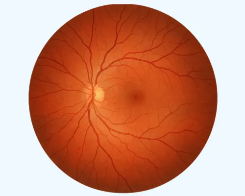
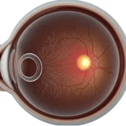
The Retina functions like a camera’s film, capturing images and sending them to the brain for interpretation. It is a light-sensitive layer located at the back of the eye. When light enters the eye, the lens focuses the image onto the retina. The retina then transforms this image into electrical signals, which travel through the optic nerve to the brain. Together with the cornea, lens, and other visual structures, the retina plays a crucial role in producing normal vision.
When the retina is damaged, it can cause significant vision problems. Difficulty in seeing objects clearly becomes the major challenge, and if left untreated, this damage may lead to permanent vision loss. Retinal surgery is performed to correct such issues, typically lasting about 1–2 hours. Since retinal diseases are among the leading causes of irreversible blindness, timely surgical intervention is critical.
Signs and Indicators of Retinal Disorders
The process of retina surgery is painless. However, there are virtually always warning signals before it develops or has progressed, such as
Floaters
Light flashes in Eyes (photopsia)

Blurred vision

Reduced side (peripheral) vision with time
Causes of Retinal Disorders
Diabetes Mellitus
Posterior vitreous detachment
Family history of retinal detachment

Trauma to your eye

Complications from cataract-removal

The strain on the eye.
Complications Linked to Retinal Disorders
In the early stages of many retinal diseases, symptoms are often absent except for slight vision changes or blurriness. As the condition progresses, vision loss may develop and worsen over time if not treated promptly and appropriately with proper medical or surgical intervention.
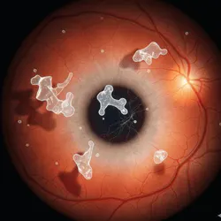
Diabetic Retinopathy
Retinopathy of Prematurity
Age-related Macular Degeneration
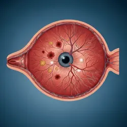
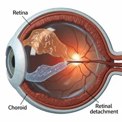
Retinal Detachment
Retina Treatment
In the initial stages of most retinal diseases, symptoms are generally absent. Over time, vision loss may occur and can worsen without timely and proper treatment. Early medical attention improves the chances of preserving or restoring vision while preventing further damage. Treatment options vary depending on the specific condition, such as diabetic retinopathy or retinal detachment, which are explained below.
Eye Injections
Retinal Laser (Photocoagulation)
Scleral buckling
Vitrectomy
Frequently Asked Questions
The retina, a crucial part of the eye, can be damaged by various disorders that may affect vision. Severe conditions such as retinal detachment, diabetic retinopathy, macular degeneration, and retinoblastoma can lead to blindness if not treated promptly.
Retinal surgery is typically completed within 1–2 hours by an eye specialist.
Yes, retina surgery is generally considered safe when performed by an experienced eye specialist. However, like any surgery, it carries some risks, such as infection, bleeding, or vision changes. Early diagnosis, proper surgical technique, and post-operative care help minimize complications.
Post-operative care and recovery time after retinal surgery can vary depending on a patient’s overall health, the type of procedure, and the severity and location of the detachment. In the first few days, mild eye pain and blurred vision are common, while swelling, redness, or tenderness may persist for several weeks.
Retinal surgery is usually not painful during the procedure because anesthesia is used. After surgery, mild discomfort, soreness, or a gritty sensation in the eye is common, but severe pain is rare. Any discomfort can typically be managed with prescribed medications.