What is Retina Surgery?
The retina functions like a camera’s film, capturing images and sending them to the brain for interpretation. It is a light-sensitive layer located at the back of the eye. When light enters the eye, the lens focuses the image onto the retina. The retina then transforms this image into electrical signals, which travel through the optic nerve to the brain. Together with the cornea, lens, and other visual structures, the retina plays a crucial role in producing normal vision.
When the retina is damaged, it can cause significant vision problems. Difficulty in seeing objects clearly becomes the major challenge, and if left untreated, this damage may lead to permanent vision loss. Retinal surgery is performed to correct such issues, typically lasting about 1–2 hours. Since retinal diseases are among the leading causes of irreversible blindness, timely surgical intervention is critical.

Symptoms of Retinal Diseases
The process of retina surgery is painless. However, there are virtually always warning signals before it develops or has progressed, such as

Floaters

Light flashes in one or both eyes (photopsia)

Blurred vision

Reduced side (peripheral) vision with time

Reduced side (peripheral) vision with time
Retinal Disease Causes

Diabetes Mellitus

Posterior vitreous detachment

Family history of retinal detachment

Trauma to your eye

Complications from cataract-removal

The strain on the eye.
Problems Associated With Retina
In the primary stages of different retinal diseases usually, no symptoms are seen alone from the one change or blurring in vision power. Later on, stages of this disorder vision losses begin to happen which may get worse if not operated timely and properly.
Diabetic Retinopathy
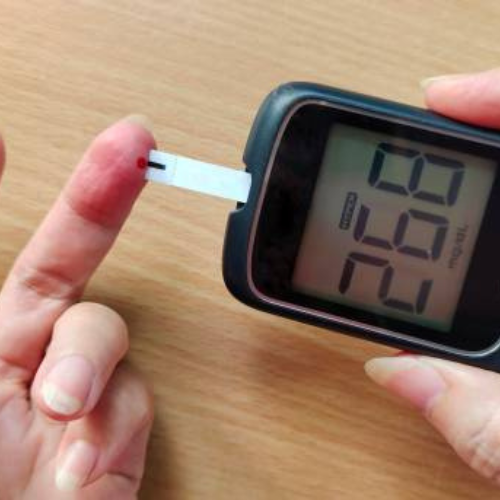
It is one of the most common issues associated with people having diabetes. In Diabetic Retinopathy disease, the high blood sugar levels cause damage to the blood vessels of the retina and damage it. In the advanced stages, it can lead to blindness.
Retinopathy of Prematurity
Retinopathy of Prematurity is mainly caused due to the retinal blood vessels developing abnormally and affects prematurely born babies. It can also lead to retinal detachment and blindness.
Age-related Macular Degeneration
The damage of the Macula can cause AMD which can lead to permanent vision loss.
Retinal Detachment
It is mainly the detachment of the Retina from the layer underneath. If it is not taken care of properly, there is a chance of a permanent vision loss. Fluorescein Angiography is one of the most common tests that are done to identify the problems which can cause permanent blindness due to the disorders of the Retina.
Retina Treatment in India
In the primary stages of different retinal diseases usually, no symptoms are seen alone. Later on, vision loss begins to happen which may get worse if not operated on timely and properly. In case of retinal disease, getting quick treatment will enhance the possibilities of retrieving or maintaining one’s vision, and limit additional loss. The treatment for retina depends on the type of disease that you are suffering from. Treatments of some of the common retinal diseases such as diabetic retinopathy, retinal detachment are discussed below:
Eye injections
These medications, called vascular endothelial protein (VEGF) inhibitors, may help stop the growth of the latest blood vessels by blocking the consequences of growth signals the body sends to get new blood vessels. Your doctor may recommend these medications, also called anti-VEGF therapy, as a stand-alone treatment or together with pan-retinal photocoagulation.
Retinal Laser (Photocoagulation)
The doctor usually uses this procedure before the stage of retinal detachment is reached i.e. only a tear or a hole. This process is called “photocococogulation”, where the doctor directs the laser into the eye through a dilated pupil, the laser causes burns around the retina, which scar and seal off the tear so that detachments do not occur. And the damage is contained.
Scleral buckling
For more severe detachments, you’ll need to have eye surgery in a hospital. Your doctor may recommend scleral buckling. This involves placing a band around the outside of your eye to push the wall of your eye into your retina, getting it back to place for correct healing. Scleral buckling may be done in combination with a vitrectomy. Cryopexy or retinopexy is performed during the scleral buckle procedure.
Vitrectomy
Another option is vitrectomy, which is used for larger tears. This procedure involves anesthesia and is often performed as an outpatient procedure, but may require an overnight stay in the hospital. Your doctor will use small tools to get rid of abnormal vascular or connective tissue and vitreous, a gel-like fluid from your retina. Then they’ll put your retina back to its proper place, commonly with a gas bubble.
